Abstract
Study of white matter at the centrum semiovale level with magnetic resonance spectroscopy and diffusion tensor imaging in cerebral small vessel disease
Author(s): L.A. Huang, X.Y. Ling, C. Li, S.J. Zhang, G.B. Chi and A.D. XuWhite matter lesion (WML) in magnetic resonance imaging is commonly observed in patients with cerebral small vessel disease (SVD), but the pathological mechanism of WML in SVD is still unclear. We observed the metabolism and microscopic anatomy of white matter in SVD patients. Twelve subjects clinically diagnosed with SVD and 6 normal control subjects were examined with magnetic resonance spectroscopy (MRS) and diffusion tensor imaging (DTI). The white matter at the centrum semiovale level was selected as the region of interest (ROI). The ROI metabolism parameters, including N-acetyl-l-aspartic acid (NAA), creatine (Cr), and choline (Cho) were measured by MRS. Microscopic parameters such as mean diffusion (MD) and fractional anisotropy (FA) in ROI were obtained by DTI. Compared with the normal control group, bilateral MD values in the SVD group were significantly elevated, whereas bilateral FA values in SVD were decreased, but the difference was not statistically significant. Additionally, NAA/Cho, Cho/Cr, and NAA/Cr showed no significant statistical differences. Our study suggests that the mechanisms of the SVD cognitive impairment are related to damage of the white matter structures rather than to brain metabolism.
Impact Factor an Index
Google scholar citation report
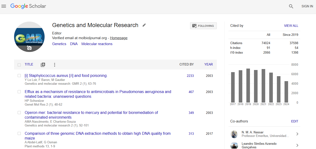
Citations : 74024
Genetics and Molecular Research received 74024 citations as per google scholar report