Abstract
Protective mechanism of quercetin on acute myocardial infarction in rats
Author(s): B. Li M. Yang J.W. Liu G.T. YinTo investigate the protective mechanism of quercetin on acute myocardial infarction (AMI) rats, an AMI rat model was established by ligating the left coronary anterior descending branch. The rats were randomly divided into the model group and low- and high-dose quercetin groups. The control group comprised sham-operated rats. The rats in the low- and high-dose quercetin groups were administered 100 and 400 mg/kg quercetin, respectively, by gavage. The rats in the control and model groups were administered isometric normal saline once daily for one week. The mRNA and protein levels of TNF-α and IL-1β in the myocardial tissue of rats were detected in each group by real time polymerase chain reaction and enzyme-linked immunosorbent assay. Malondialdehyde (MDA) content in the myocardial tissue and superoxide dismutase (SOD) and catalase (CAT) activities were detected using a colorimetric method. The level of apoptosis was detected by terminal deoxynucleotidyl transferase dUTP nick end labeling. Compared with those in the control group, the mRNA and protein levels of TNF-α, IL-1β and MDA content in the model, low-, and high-dose groups significantly increased. SOD and CAT activities decreased significantly. The cell apoptosis index increased significantly (P To investigate the protective mechanism of quercetin on acute myocardial infarction (AMI) rats, an AMI rat model was established by ligating the left coronary anterior descending branch. The rats were randomly divided into the model group and low- and high-dose quercetin groups. The control group comprised sham-operated rats. The rats in the low- and high-dose quercetin groups were administered 100 and 400 mg/kg quercetin, respectively, by gavage. The rats in the control and model groups were administered isometric normal saline once daily for one week. The mRNA and protein levels of TNF-α and IL-1β in the myocardial tissue of rats were detected in each group by real time polymerase chain reaction and enzyme-linked immunosorbent assay. Malondialdehyde (MDA) content in the myocardial tissue and superoxide dismutase (SOD) and catalase (CAT) activities were detected using a colorimetric method. The level of apoptosis was detected by terminal deoxynucleotidyl transferase dUTP nick end labeling. Compared with those in the control group, the mRNA and protein levels of TNF-α, IL-1β and MDA content in the model, low-, and high-dose groups significantly increased. SOD and CAT activities decreased significantly. The cell apoptosis index increased significantly (P
Impact Factor an Index
Google scholar citation report
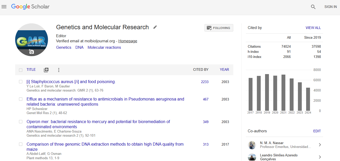
Citations : 74024
Genetics and Molecular Research received 74024 citations as per google scholar report