Abstract
Mechanism of rat osteosarcoma cell apoptosis induced by a combination of low-intensity ultrasound and 5-aminolevulinic acid in vitro
Author(s): Y.N. Li, Q. Zhou, B. Yang, Z. Hu, J.H. Wang, Q.S. Li and W.W. CaoWe investigated the killing effect of low-intensity ultrasound combined with 5-aminolevulinic acid (5-ALA) on the rat osteosarcoma cell line UMR-106. Logarithmic-phase UMR-106 cells were divided into a control group, ultrasound group and 5-ALA group. The cell apoptotic rate, production of reactive oxygen species, and the change in mitochondrial membrane potential were analyzed by flow cytometry; ultrastructural changes were observed by transmission electron microscopy. Using low-intensity ultrasound at 1.0 MHz and 2.0 W/cm2 plus 5-ALA at a concentration of 2 mM, the apoptotic rate of the sonodynamic therapy group was 27.2 ± 3.4% which was significantly higher than that of the control group, ultrasound group, and 5-ALA group (P < 0.05). Theproduction of reactive oxygen species was 32.6 ± 2.2% and the decrease in mitochondrial membrane potential was 39.5 ± 2.5%. The 33342 staining showed nuclear condensation and fragmentation in the ultrasound group and 5-ALA group. Structural changes in the cell membrane, mitochondria, Golgi apparatus, and other organelles observed by transmission electron microscopy included formation of apoptotic bodies. The killing effect of low-intensity ultrasound combined with 5-ALA on UMR-106 cells was significant. Cell apoptosis played a vital role in the killing effect, and the mitochondria pathway contributed to the apoptosis of UMR-106 cells.
Impact Factor an Index

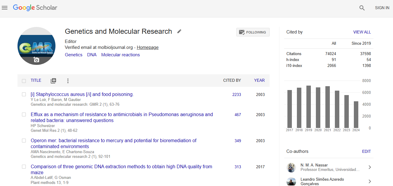
Google scholar citation report
Citations : 74024
Genetics and Molecular Research received 74024 citations as per google scholar report
