Abstract
Isolation and characterization of white and brown adipocytes in Kunming mice
Author(s): Y.W. Nie*, P. Zhang*, J. Zhang, H.Y. Liang, M. Wang, B. Dai, H. Liangand D.J. LiuWhite adipose tissue and brown adipose tissue play critical roles in controlling energy homeostasis and the development of obesity and diabetes. We isolated mouse white adipocytes from inguinal white fat tissues and brown adipocytes from interscapular brown fat tissues, and employed a variety of approaches, including immunofluorescent staining, quantitative real-time PCR, western blotting analysis, and differentiation assay, to characterize those adipocytes. Both white and brown adipocytes stained positively for CD44 and CD29, and lipid droplets were observed after the induction of adipogenesis. The Asc1 expression level in the white adipocytes was 2.5-fold higher than that in the brown adipocytes (P Cidea mRNA level (P White adipose tissue and brown adipose tissue play critical roles in controlling energy homeostasis and the development of obesity and diabetes. We isolated mouse white adipocytes from inguinal white fat tissues and brown adipocytes from interscapular brown fat tissues, and employed a variety of approaches, including immunofluorescent staining, quantitative real-time PCR, western blotting analysis, and differentiation assay, to characterize those adipocytes. Both white and brown adipocytes stained positively for CD44 and CD29, and lipid droplets were observed after the induction of adipogenesis. The Asc1 expression level in the white adipocytes was 2.5-fold higher than that in the brown adipocytes (P Cidea mRNA level (P
Impact Factor an Index
Google scholar citation report
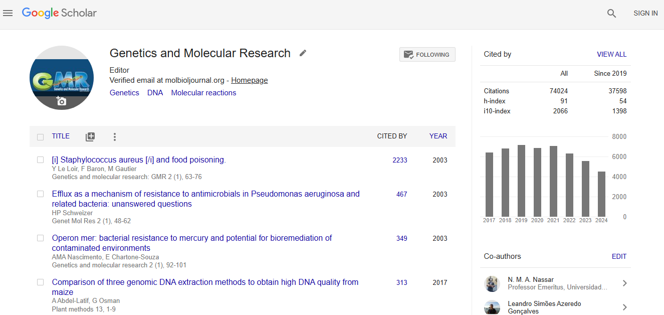
Citations : 74024
Genetics and Molecular Research received 74024 citations as per google scholar report