Abstract
Inhibitory effects of spironolactone on myocardial fibrosis in spontaneously hypertensive rats
Author(s): H. Zhao, D.W. Gu, H.T. Li, Q.F. Ge and G.P. LiThis study evaluated the inhibitory effects of spironolactone, a non-selective aldosterone receptor antagonist, on hypertension-induced myocardial fibrosis. Collagen I and III contents was detected in the myocardial tissue of spontaneously hypertensive rats (SHRs) after spironolactone administration. Twenty male SHRs were assigned to the spironolactone group or control group (N = 10 each); 7 Wistar-Kyoto rats (WKY) were also used. Spironolactone dissolved in ddH2O was administered via gavage at a dosage of 20 mg·kg-1·day-1. Meanwhile, the control and WKY groups were administered equivalent volumes of ddH2O for 16 weeks. Western blotting was used to detect the contents of collagen I in myocardial tissue; observations were performed using polarizing microscopy, and the area integration and ratio of collagen I/III were subsequently calculated. Compared to the WKY group, collagen I synthesis was significantly higher in the control group (1.87 ± 0.2 vs 1.21 ± 0.7, P < 0.05). After 16 weeks of treatment, collagen I contents were significantly lower in the spironolactone group than in the control group (1.42 ± 0.05 vs 1.87 ± 0.2, P < 0.05). The areas of collagen I and collagen I/III ratio were significantly smaller in the spironolactone group than in the control group (6400 ± 259 vs 12,019 ± 734 pixels, 15.64 ± 1.34 vs 20.8 ± 3.04 pixels, respectively; P < 0.05). However, there were no significant differences in the area of collagen III among the three groups. In conclusion, spironolactone improves myocardial collagen deposition, preventing myocardial fibrosis in SHRs.
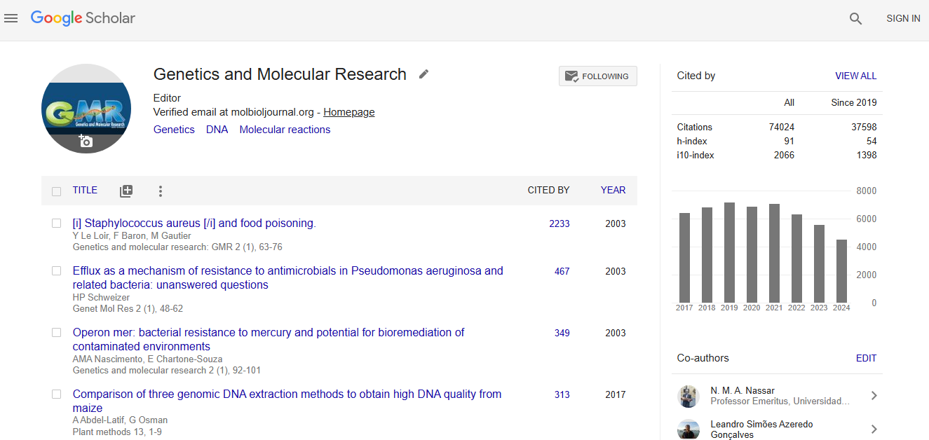
Impact Factor an Index
Google scholar citation report
Citations : 74024
Genetics and Molecular Research received 74024 citations as per google scholar report