Abstract
Evaluation of bone matrix gelatin/fibrin glue and chitosan/gelatin composite scaffolds for cartilage tissue engineering
Author(s): Z.H. Wang, J. Zhang, Q. Zhang, Y. Gao, J. Yan, X.Y. Zhao, Y.Y. Yang, D.M. Kong, J. Zhao, Y.X. Shi and X.L. LiThis study was designed to evaluate bone matrix gelatin (BMG)/fibrin glue and chitosan/gelatin composite scaffolds for cartilage tissue engineering. Chondrocytes were isolated from costal cartilage of Sprague-Dawley rats and seeded on BMG/fibrin glue or chitosan/gelatin composite scaffolds. After different in vitro culture durations, the scaffolds were subjected to hematoxylin and eosin, Masson’s trichrome, and toluidine blue staining, anti-collagen II and anti-aggrecan immunohistochemistry, and scanning electronic microscopy (SEM) analysis. After 2 weeks of culture, chondrocytes were distributed evenly on the surfaces of both scaffolds. Cell numbers and the presence of extracellular matrixcomponents were markedly increased after 8 weeks of culture, and to a greater extent on the chitosan/gelatin scaffold. The BMG/fibrin glue scaffold showed signs of degradation after 8 weeks. Immunofluorescence analysis confirmed higher levels of collagen II and aggrecan using the chitosan/gelatin scaffold. SEM revealed that the majority of cells on the surface of the BMG/fibrin glue scaffold demonstrated a round morphology, while those in the chitosan/gelatin group had a spindle-like shape, with pseudopodia. Chitosan/gelatin scaffolds appear to be superior to BMG/ fibrin glue constructs in supporting chondrocyte attachment, proliferation, and biosynthesis of cartilaginous matrix components.
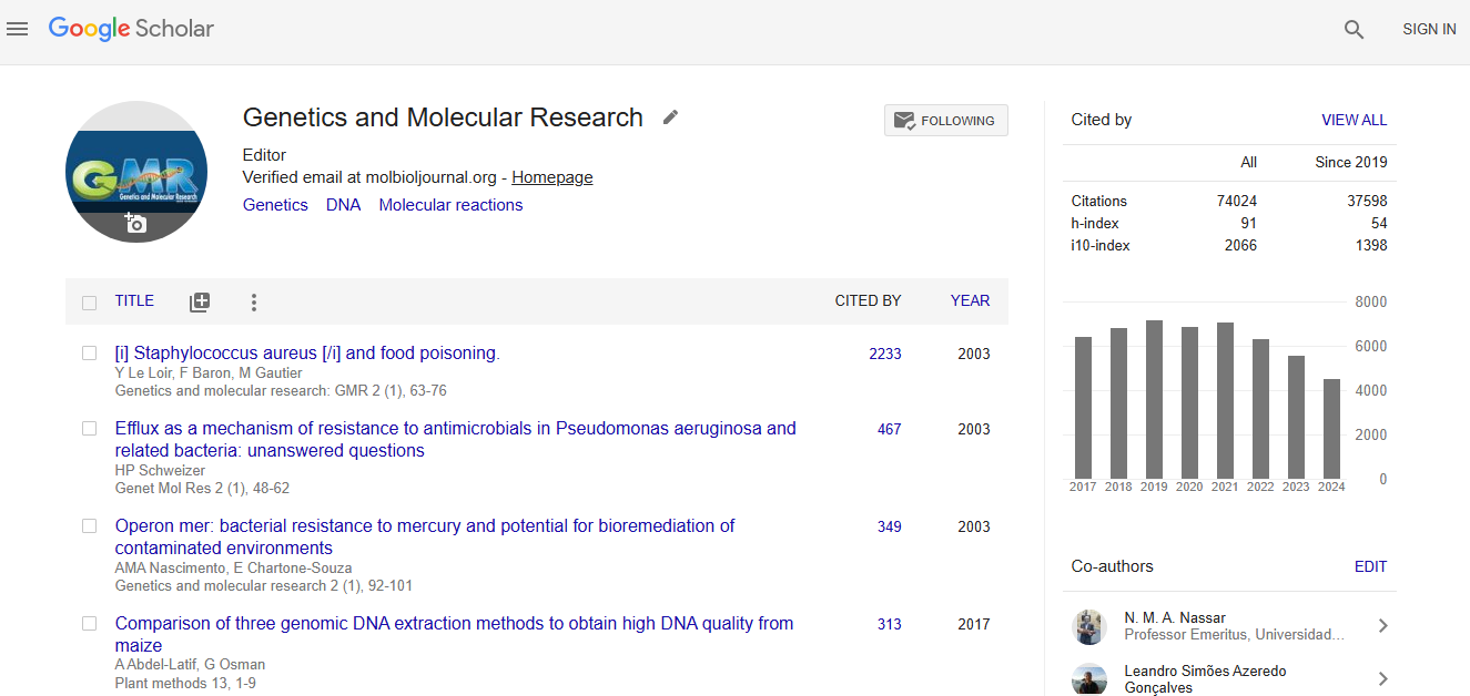
Impact Factor an Index
Google scholar citation report
Citations : 74024
Genetics and Molecular Research received 74024 citations as per google scholar report