Abstract
Correlation between magnetic resonance perfusion weighted imaging of radiation brain injury and pathology
Author(s): X.J. Liu, C.F. Duan, W.W. Fu, L. Niu, Y. Li, Q.L. Sui and W.J. XuWe used magnetic resonance perfusion weighted imaging and pathological evaluation to examine different stages of radiation-induced brain injury and to investigate the correlation between the relative cerebral blood volume (rCBV) ratio and vascular endothelial growth factor (VEGF). Thirty adult rats were randomly divided into 2 groups: control and radiation group. The control group was not subjected to irradiation. The irradiation group rats were examined by magnetic resonance imaging and magnetic resonance perfusion weighted imaging at 1, 3, 6, 9, and 12 months after radiation treatment. We measured the rCBV, mean transit time, and time to peak. Hematoxylin and eosin staining, immunohistochemical staining, and electron microscopy were performed. VEGF absorbance was evaluated by immunohistochemical staining. Compared with the control group, the differences in rCBV, mean transit time, time to peak, and VEGF absorbance after 3 months were statistically significant (P < 0.05). rCBV was positively correlated with VEGF (r = 0.94, P < 0.05).
Impact Factor an Index

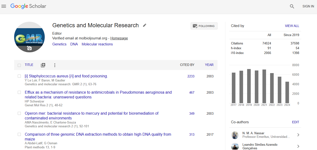
Google scholar citation report
Citations : 74024
Genetics and Molecular Research received 74024 citations as per google scholar report
