Abstract
Comparative Study of Blood-Derived Scaffolds for the Culture of Human Adipose Derived Stem Cells (ASCS) and Dermal Fibroblasts
Author(s): Helga Caputo Nunes, Rosana Rossi Ferreira,Ana Carolina Picolo Pasian, Andrei Moroz, Bruno Martinucci, Michele Janegitz Acorci Valerio,S√?¬©rgio Alexandre Alc√?¬¢ntara dos Santos, Henrique de Souza Vieira,S√?¬©rgio Luis Felisbino, Elenice Deffune, Fl√?¬°via Karina DelellaThis study aimed to compare the performance of bloodderived scaffolds with a well-known and accepted scaffold, chitosan, in maintaining cell cultures of ASCs and fibroblasts for future wound healing applications. Cells were characterized, immunophenotyped and cultivated into the following scaffolds: 1) Chitosan (CH, control), 2) Platelet gel (PG), 3) Chitosan blended with platelet-derived growth factors (CHPG), and 4) Fibrin glue (FG). Parameters were evaluated: i) Maintenance of cell morphology ii) Cell proliferation and iii) Citotoxicity. Our results show that ASCs and fibroblasts presented similar proliferation behaviors, which were scaffold-dependent. Regarding cell density, there were more cells in PG, followed by CHPG, FG, and CH scaffolds, for both cell types. Moreover, apoptosis assays revealed that CH had the highest rates of early (4.7%) and late apoptosis (13.9%). The proposed scaffolds demonstrated significantly lower levels of apoptosis, at less than 10%. For all these reasons, our findings demonstrate that when compared to CH, both ASCs and fibroblasts may be grown more efficiently in all three proposed scaffolds. Furthermore, we can also conclude that PG and CHPG seems to be the better choices as biomaterials for the expansion of these cells, due to higher cell proliferation, and lower apoptosis levels. Finally, it is possible to conclude that a surplus from blood bank components may be used as scaffolds with bioactive properties, providing a suitable microenvironment for cells, which could then be employed to establish tissue banks for wound healing applications.
Impact Factor an Index
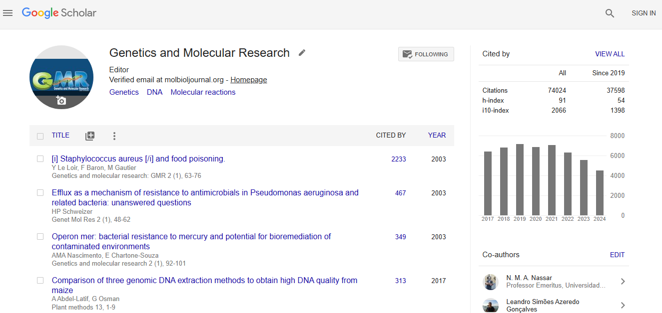
Google scholar citation report
Citations : 74024
Genetics and Molecular Research received 74024 citations as per google scholar report