Abstract
Characterization of abnormal epithelium after laser-assisted subepithelial keratectomy using in vivo confocal microscopy
Author(s): J. Zuo, C.W. Zhang, X. Zhou, W. Wei and Y.L. WangThis study compared the abnormal corneal epithelium after laser-assisted subepithelial keratectomy (LASEK) with dystrophic cornea using in vivo using confocal microscopy (IVCM) and examined the effects of the abnormal epithelium on postoperative recovery of uncorrected distance visual acuity (UDVA) and sub-basal nerve plexus regeneration. The UDVA and wound healing were examined in 50 eyes of 25 patients undergoing LASEK at 1 week, 1 month, and 1 year postoperatively, including the visual acuity, slit lamp microscopy, and IVCM. After 1 week, the corneal epithelium was healed in all patients, but abnormal epithelial cells were detected in 33/50 patients using IVCM. Abnormal cells were limited to the surgical margin, and highly reflective granules were observed underneath. At 1 month and 1 year postoperatively, the abnormal epithelium was unchanged in size. At 1 year postoperatively, there were clear differences between the sub-basal nerve plexus in the normal and abnormal epithelium. At 1month postoperatively, the UDVA was >1.0 in 88% of patients, which increased to 94% at 1 year, and there was no clear difference in the UDVA between abnormal (N = 33) and normal (N = 17) epithelium. After LASEK, abnormal epithelial cells may arise at the margin of the epithelial flap and persist 1 year postoperatively accompanied by poor regeneration of the sub-basal nerve plexus. However, this does not affect the UDVA postoperatively. The abnormal epithelium may be caused by residual necrotic basal cell debris on the epithelial basement membrane and abnormal neurotrophic metabolism between the corneal epithelium and nerve plexus.
Impact Factor an Index
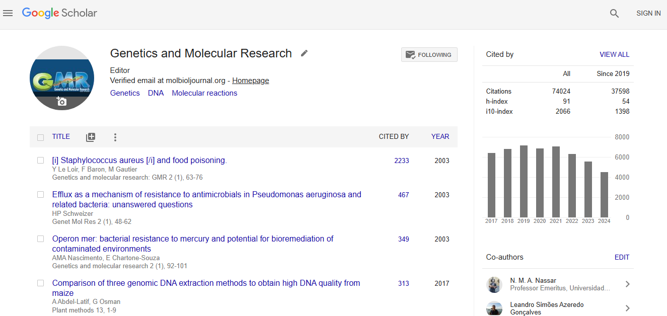
Google scholar citation report
Citations : 74024
Genetics and Molecular Research received 74024 citations as per google scholar report